Tuesday, August 28, 2007

REFLEX ACTION

REFLEX ACTION
A reflex action is a coordinated response to a specific stimulus.

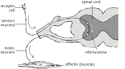
The following figure shows a reflex arc:


The sequence of events in a reflex action (Example: touching hot object):
  • The sensory receptor in the skin first received a stimulus (hot object).
  • This caused an electrical impulse to be generated which is then transmitted by the sensory neurones towards the spinal cord.
  • In the spinal cord, at the synapse between the sensory neurones and the relay neurones (at the nerve endings of the sensory neurones) a chemical called acetylcholine, is released which stimulates the relay neurones to produce an electrical impulse (nerve impulse). The nerve impulse is then transmitted along the relay neurones towards the motor neurones.
  • At the synapse between the relay neurones and the motor neurones (at the nerve endings of the relay neurones) acetylcholine is again released which stimulates the motor neurones to produce an electrical impulse.
  • This electrical impulse is then transmitted along the motor neurones towards the effectors (in this case the effectors are the biceps and triceps of the arm).
  • A response is then produced as the biceps contracts and the triceps relaxes. This antagonistic action of the biceps and triceps helps to lift the hand away from the hot object.

OSMOREGULATION

OSMOREGULATION: REGULATION OF BLOOD WATER POTENTIAL

DECREASE IN BLOOD WATER POTENTIAL
  • Decrease may be due to loss of water through sweating when doing vigorous exercise or when the environmental temperature increases which causes more sweating (stimulus).
  • This decrease stimulates the pituitary gland in the brain to secrete more Anti Diuretic Hormone (ADH) into the blood.
  • The blood then transports this ADH to the kidneys (target organ).
  • The function of ADH is to promote water reabsorption from the filtrate in the kidney tubules back into the blood capillaries which are in close contact with the kidney tubules.
  • Since ADH is secreted in high concentration, more water will be reabsorbed back from the filtrate in the kidney tubules into the blood capillaries.
  • As a result, urine production will decrease and hence urine becomes more concentrated.
  • Reabsorption of water back into the blood capillaries, causes the water potential in the blood to increase (Negative Feedback) and eventually the level of water potential in the blood will return back to normal.
(Note: if this corrective mechanism is not functioning, red blood cells in the blood may undergo crenation and hence less oxygen will be transported)

INCREASE IN BLOOD WATER POTENTIAL
  • Increase may be due to too much intake of water or lack of sweating (stimulus).
  • This increase stimulates the pituitary gland in the brain to secrete less ADH into the blood.
  • The blood then transports this ADH to the kidneys (target organ).
  • The function of ADH is to promote water reabsorption from the filtrate in the kidney tubules back into the blood capillaries which are in close contact with the kidney tubules.
  • Since ADH is secreted in low concentration, less water will be reabsorbed back from the filtrate in the kidney tubules into the blood capillaries.
  • As a result, urine production will increase and hence urine becomes more dilute.
  • Less reabsorption of water back into the blood capillaries, causes the water potential in the blood to decrease (Negative Feedback) and eventually the level of water potential in the blood will return back to normal.

TEMPERATURE REGULATION

INCREASE IN BODY TEMPERATURE
  • An increase in body temperature, for example due to vigorous exercise or being in a warm place, causes the blood and the skin temperature to increase (stimulus).
  • The temperature receptors in the skin detect this increase.
  • A signal in the form of nerve impulse is then transmitted to the hypothalamus in the brain.
  • When the hypothalamus is stimulated, it then transmits nerve impulses to the relevant body parts.
  • The arterioles in the skin dilate. More blood is transported to the blood capillaries and this removes more heat.
  • Sweat glands become active and more sweating occurs and hence more heat removed.
  • The hair erector muscles in the skin relax causing the hair to lie flat. Therefore less heat is trapped by the hair.
  • Metabolic rate decreases and hence heat production decreases.
  • Breathing rate increases so as to remove more heat in exhaled air.
  • All these mechanisms lower down the blood temperature (Negative feedback) and eventually the temperature will return to normal.
DECREASE IN BODY TEMPERATURE
  • A decrease in body temperature, for example due to starvation, fasting or being in cold places, causes the blood and the skin temperature to decrease (stimulus).
  • The temperature receptors in the skin detect this decrease.
  • A signal in the form of nerve impulse is then transmitted to the hypothalamus in the brain.
  • When the hypothalamus is stimulated, it then transmits nerve impulses to the relevant body parts.
  • The arterioles in the skin constrict. Less blood is transported to the blood capillaries and hence less heat is removed.
  • Sweat glands become inactive and no sweating occurs. Therefore no heat is removed.
  • The hair erector muscles in the skin contract causing the hair to stand erect. Therefore more heat is trapped by the hair. (Remember: still air trapped in between the hairs acts as an insulator, therefore less heat is removed)
  • Metabolic rate increases and hence heat production increases.
  • Breathing rate decreases so as to conserve more heat.
  • Shivering due to muscle contractions occurs if very cold to produce more heat.
  • All these mechanisms increases the blood temperature (Negative feedback) and eventually the temperature will return to normal.

REGULATION OF GLUCOSE CONCENTRATION

REGULATION OF BLOOD GLUCOSE CONCENTRATION

What happens if the glucose concentration in the blood increases?
  • After having a meal, the glucose concentration in the blood increases (stimulus).
  • This increase may caused cells in the body to undergo crenation. For example, the red blood cells in the blood may undergo crenation due to higher water potential in the red blood cells than the blood itself. As a result water molecules in the red blood cells diffuse out of the red blood cells by osmosis.
  • This condition needs to be corrected. Because of this increase, the pancreas will secrete insulin into the blood (insulin is a hormone produced by the beta-cells of the islets of langerhans of the pancreas).
  • The blood will then transports the insulin to the liver (target organ).
  • In the liver, the excess glucose is then converted into glycogen for storage in the liver itself as well as in the muscles.
  • This conversion of excess glucose to glycogen slowly lowers the blood glucose concentration (negative feedback).
  • Eventually the glucose concentration in the blood will return to normal (Norm).
What happens if the glucose concentration in the blood decreases?
  • After vigorous exercise, the glucose concentration in the blood decreases due to glucose being oxidised to release energy during cellular respiration (stimulus)
  • The decreased may caused the red blood cells to haemolyse. Haemolysis occurs due to passage of water molecules into the red blood cells from the blood from higher to lower water potential by osmosis.
  • This condition needs to be corrected. The pancreas responds to this by producing glucagon into the blood (Glucagon is a hormone produced by the alpha cells of the islets of langerhans of the pancreas).
  • The glucagon is then transported by the blood to the liver which is the target organ.
  • In the liver, the glucagon converts glycogen stored in the liver into glucose.
  • This will boost the blood glucose concentration in the blood (negative feedback)
  • Eventually the blood glucose concentration will return to normal.
Note: Before the insulin and the glucagon can be secreted into the blood, the pancreas must first be triggered or stimulated by the pituitary gland in the brain (Remember: Pituitary gland is a MASTER gland of all the endocrine glands. It controls the activities of other endocrine glands)

THE SKIN

THE ROLE OF THE SKIN IN PREVENTING OVERHEATING
Overheating occurs when too much heat is being produced by the body so the idea here is to remove the excess heat. This then prevent the body temperature to rise above normal. The following are the mechanisms involved in preventing overheating (all occurs within the skin itself):
  • The arterioles in the skin vaso-dilate (get wider). As a result of this more blood is directed or transported to the blood capillaries near to the surface of the skin. By this, heat is brought near to the surface of the skin.
  • The sweat glands make more sweat. The sweat is then deposited through the sweat pore to the surface of the skin. The heat from the blood is used to evaporate the sweat and at the same time when the sweat evaporates, heat is also removed at the same time.
  • The hair erector muscles relax and as a result the body hairs lie flat against the skin allowing air current to get nearer to the skin. As a result more heat will be removed.
(Apart from what happens in the skin the following also occur in order to remove more heat from the body)
  • Cellular metabolism in the cells slows down so less heat is produced.
  • Breathing rate increases so more heat is removed with the expired air.

THE ROLE OF THE SKIN IN PREVENTING OVERCOOLING
Overcooling occurs when not enough heat is being produced by the body. Therefore the body tries to conserve heat as much as possible so as to prevent the body temperature to drop below normal. The following mechanisms are very important in preventing overcooling:
  • The arterioles in the skin vaso-constrict (becomes narrower) and as a result less blood is transported or directed to the blood capillaries. This prevent heat being removed.
  • Sweat productions stops or no sweating occurs. Therefore no heat removed.
  • The hair erector muscles contract raising the body hairs. This prevents air current from getting near the skin surface when air is trapped in between the hairs (Note: still air is a bad conductor of heat). This prevents heat from being removed.
(Apart from the skin, the following are also involved in conserving the heat available in the body as well as in preventing heat loss)
  • Cellular metabolism increases, producing more heat for the blood to distribute around the body.
  • The skeletal muscles start contracting regularly which causes shivering. Shivering generates heat.
HYPERTHERMIA AND HYPOTHERMIA
  • Hypothermia is a condition where the body temperature falls below normal because too little heat is being generated.
  • Hyperthermia is a condition which occurs when the body is unable to shed enough heat to stop the temperature from rising.

THE BRAIN

Central Nervous System

The Central Nervous System (CNS) is composed of the brain and spinal cord. The CNS is surrounded by bone-skull and vertebrae. Fluid and tissue also insulate the brain and spinal cord.

The brain is composed of three parts: the cerebrum (seat of consciousness), the cerebellum, and the medulla oblongata (these latter two are "part of the unconscious brain").

The medulla oblongata is closest to the spinal cord, and is involved with the regulation of heartbeat, breathing, vaso-constriction (blood pressure), and reflex centers for vomiting, coughing, sneezing, swallowing, and hiccuping. The hypothalamus regulates homeostasis. It has regulatory areas for thirst, hunger, body temperature, water balance, and blood pressure, and links the Nervous System to the Endocrine System. The midbrain and pons are also part of the unconscious brain. The thalamus serves as a central relay point for incoming nervous messages.

The cerebellum is the second largest part of the brain, after the cerebrum. It functions for muscle coordination and maintains normal muscle tone and posture. The cerebellum coordinates balance.

The conscious brain includes the cerebral hemispheres. The cerebrum governs intelligence and reasoning, learning and memory. While the cause of memory is not yet definitely known, studies on slugs indicate learning is accompanied by a synapse decrease. Within the cell, learning involves change in gene regulation and increased ability to secrete transmitters.

The Medulla Oblongata and the Midbrain

The medulla oblongata controls heart rate, constriction of blood vessels, digestion and respiration.

The midbrain consists of connections between the hindbrain and forebrain. Mammals use this part of the brain only for eye reflexes.

The Cerebellum

The cerebellum is the third part of the hindbrain, but it is not considered part of the brain stem. Functions of the cerebellum include fine motor coordination and body movement, posture, and balance. This region of the brain is enlarged in birds and controls muscle action needed for flight.

The Forebrain

The forebrain consists of the diencephalon and cerebrum. The thalamus and hypothalamus are the parts of the diencephalon. The thalamus acts as a switching center for nerve messages. The hypothalamus is a major homeostatic center having both nervous and endocrine functions.

The cerebrum, the largest part of the human brain, is divided into left and right hemispheres connected to each other by the corpus callosum. The hemispheres are covered by a thin layer of gray matter known as the cerebral cortex.
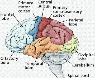
The cortex in each hemisphere of the cerebrum is between 1 and 4 mm thick. Folds divide the cortex into four lobes: occipital, temporal, parietal, and frontal. No region of the brain functions alone, although major functions of various parts of the lobes have been determined.

The occipital lobe (back of the head) receives and processes visual information. The temporal lobe receives auditory signals, processing language and the meaning of words. The parietal lobe is associated with the sensory cortex and processes information about touch, taste, pressure, pain, and heat and cold. The frontal lobe conducts three functions:

  1. motor activity and integration of muscle activity
  2. speech
  3. thought processes

Most people who have been studied have their language and speech areas on the left hemisphere of their brain. Language comprehension is found in Wernicke's area. Speaking ability is in Broca's area. Damage to Broca's area causes speech impairment but not impairment of language comprehension. Lesions in Wernicke's area impairs ability to comprehend written and spoken words but not speech. The remaining parts of the cortex are associated with higher thought processes, planning, memory, personality and other human activities.

The Spinal Cord

The spinal cord runs along the dorsal side of the body and links the brain to the rest of the body. Vertebrates have their spinal cords encased in a series of (usually) bony vertebrae that comprise the vertebral column.

The gray matter of the spinal cord consists mostly of cell bodies and dendrites. The surrounding white matter is made up of bundles of interneuronal axons (tracts). Some tracts are ascending (carrying messages to the brain), others are descending (carrying messages from the brain). The spinal cord is also involved in reflexes that do not immediately involve the brain.
HORMONES

WHAT ARE HORMONES?
Hormones are chemical substances produced in minute quantities by the endocrine glands and transported by the blood to the target organs where they exert a profound effect. After exerting its effect in the target organ, it is then destroyed in the liver and then transported to the kidneys to be excreted.

DIFFERENCES BETWEEN NERVOUS CONTROL AND HORMONAL CONTROL
  • NC involves nervous impulses whereas HC involves hormones
  • Impulses are transmitted by neurones in NC whereas hormones are transmitted by blood in HC
  • The response is very quick in NC and slower in HC
  • Response in NC is usually short-lived whereas in HC it may be short-lived or long-lived
  • NC may be voluntary or involuntary whereas HC is always involuntary
  • NC is usually localised and in HC it may affects more than one target organ

ADRENALINE
  • Adrenaline is a hormone produced by the Adrenal gland located just above each kidney
  • Circumstances in which it is secreted: Usually in conditions of fear, anger and anxiety or in any other emergency situation
  • Effect 1: Increases metabolic rate. This means that more energy is released in tissue respiration
  • Effect 2: Increases in rate of heartbeat and rise in blood pressure so that oxygen and glucose are carried faster to the muscles
  • Effect 3: Constriction of arterioles in skin which causes pallor. As a result of this more blood is sent to the muscles
  • Effect 4: Causes the conversion of more glycogen stored in the liver into glucose so that the glucose can be used in tissue respiration
  • Effect 5: Dilates the pupil of the eye so that more light can enter the eye. Hence this provides clearer vision
  • Effect 6: Hair muscles may contract producing "goose pimples"
INSULIN
  • Insulin is a hormone produced by the Islets of Langerhans in the pancreas
  • Secretion of insulin is due to an increase in concentration of glucose in the blood
  • Effect 1: Causes the conversion of glucose to glycogen for storage in the liver and muscles
  • Effect 2: Enables tissue cells to oxidise glucose to produce energy in tissue respiration (Remember the "Gate Keeper")
  • Both effects help to control the amount of glucose in the blood

THE HUMAN EYES

1. PARTS AND FUNCTIONS


  • Sclera - Tough white outer coating (the white part of your eye is actually the sclera. The main function of the sclera is to protect the eyeball.
  • Cornea - This is actually the front part of the sclera but unlike the sclera it is transparent. The function of the cornea is to refract light rays into the eye.
  • Conjunctiva - This is a thin epithelium which protect the cornea.
  • Vitreous humour and aqueous humour - The liquid behind the lens is jelly-like and is called vitreous humour while the aqueous humour in front of the lens is watery. The function of both the vitreous humour and the aqueous humour is to keep the spherical shape of the eyeball. In addition, the aqueous humour also functions in providing nourishment to the non-vascularised lens and cornea.
  • Lens - Transparent structure (flexible and can change its shape during accomodation) which refract light rays on to the retina.
  • Suspensory ligament - To hold the lens in place. Plays a very important role in accomodation.
  • Iris - Iris gives colour to your eyes. It consists of the radial and the circular muscles. The iris controls the size of the pupil, thus controlling the amount of light entering the eyes. These muscles of the iris work antagonistically.
  • Pupil - Pupil is a hole at the centre of the iris. The function is to allow light to enter the eye.
  • Choroid - The second layer of the eye. It is highly vascularised to provide nourishment to the eye. It is also pigmented black (absorbs light) so as to prevent internal reflection in the eye.
  • Ciliary body - It produces aqueous humour. It contains circular muscles which helps to alter the size of the lens during accomodation.
  • Retina - The internal lining at the back of the eye is the retina. It contains light sensitive cells (the cones and the rods) which respond to light.
  • Fovea (yellow spot) - The part of the retina which is very sensitive to light since rods and cones are highly concentrated here. When you focus on an object, the image of the object will fall on to this region.
  • Blind spot - This region of the retina contains no light sensitive cells so object will not be seen here if light falls on to this region.
  • Optic nerve - This contains nerve fibres which transmit electrical impulses to the brain.
  • Tear glands - These are glands present under the top of the eyelid. The function is to produce tear fluid which helps to prevent friction when blinking. The tear fluid also wash away any dust particles or foreign bodies. It contains lysozyme which kills bacteria.

2. ACCOMODATION


  • Viewing distant image - Ciliary muscles relax, the suspensory ligaments become taut pulling the lens outwards. As a result the lens becomes thinner. This will in turn, increased the focal length hence enable you to view distant object.
  • Viewing near object - Ciliary muscles contract, the suspensory ligaments become slacken. Hence there is no pulling force to pull the lens outwards. As a result the lens becomes thicker and this will decrease the focal length. Hence you are able to focus at near object.

3. PUPIL REFLEX

The following figure shows the iris which control the size of the pupil thus controlling the amount of light entering the eye.


In Bright Light AND In Dim Light
  • The retina (due to the presence of rods and cones) is very sensitive to light. When light falls on to the retina, the light (stimulus) will stimulates the retina. An electrical impulse will then be transmitted to the brain along the sensory nerve fibres in the optic nerve which contains sensory neurones. At the synapse between the sensory neurones and the relay neurones, acetylcholine will be released which stimulates the relay neurones in the brain to produce an electrical impulse. The electrical impulse is then transmitted along the relay neurones. Again at the synapse between the relay neurones and the motor neurones, acetylcholine will be released which stimulates the motor neurones to produce an electrical impulse. The electrical impulse is then transmitted along the motor neurones to the effector (which in this case are the radial and circular muscles of the iris).
  • In dim light, the radial muscles contract and the circular muscles relax. This causes the pupil to dilate and hence allows more light to enter the eye.
  • In bright light, the radial muscles relax and the circular muscles contract. This causes the pupil to constrict and hence allows less light to enter the eye (thus protects the delicate light sensitive cells in the retina)
Note: TIME WAITS FOR NO MAN

SKELETON

FUNCTIONS OF THE SKELETON:
  • Support - The skeleton holds the body off the ground and keeps its shape even when muscles are contracting to produce movement.
  • Protection - The brain is protected from injury by being enclosed in the skull. The heart, lungs and liver are protected by the rib cage and the spinal cord is enclosed inside the backbone.
  • Movement and Locomotion - Many bones of the skeleton acts as levers. When muscles pull on these bones, they produce movements such as the raising of the ribs during breathing or the chewing action of the jaws. For a skeletal muscle to produce movement, both its ends need to have a firm attachment. The skeleton provides suitable points of attachment for the ends of muscles. The skeleton with the help of muscles also helps to move the body from one place to another. This is called locomotion.
  • Production of blood cells - The red bone marrow of some bones produce both red and white blood cells.
Note: You should be able to identify the humerus, ulna, radius, femur, tibia, fibula, pelvic girdle, pectoral girdle and of course the skull, ribs and rib cage, sternum and skull.JOINTS:
  • Where two bones meet they form a joint.
  • In the syllabus, you only have to know the hinge and the ball and socket joints.
THE HINGE JOINT

THE BALL AND SOCKET JOINT
  • Where can these joints be found? Hinge joints are found at your elbows and your knees whereas the ball and socket joint are usually found at your shoulder and your hip.
  • The differences between these two types of joints are: hinge joints only allow movement in one direction only (for example bending your knee and your arm) whereas the ball and socket joints allow movement in all direction.

THE CARTILAGE, LIGAMENT, SYNOVIAL MEMBRANE AND SYNOVIAL FLUID

  • The cartilage functions in reducing friction between two bones as shown in the figure above.
  • Friction is further reduced by the synovial fluid which is produced by the synovial membrane.
  • The function of the ligaments is to hold bones in position thus preventing dislocation. Dislocation may occurs if the ligaments are torn.
ANTAGONISTIC ACTION OF THE BICEPS AND TRICEPS IN THE ARM
  • You need to know the role the biceps and triceps (these are known as skeletal muscles) in your arm which helps in bringing about movement such as when you are flexing or stretching your arm.
  • Arm flexing - Biceps contracts while triceps relaxes.
  • Arm stretching - Biceps relaxes while triceps contracts.